From Traditional Scans to Advanced Radiological Technologies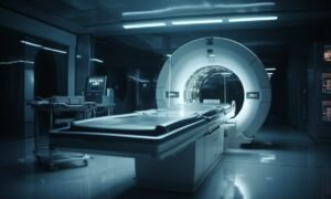

The Evolution of Diagnostic Imaging
Diagnostic imaging has developed through three stages which started with basic imaging methods and progressed to advanced technology systems that provide accurate medical assessments.
The field of radiological research began with basic radiographic methods, but now includes advanced radiological systems which combine physics, computing, and artificial intelligence to improve patient outcomes.
Today, diagnostic imaging serves as both a disease detection tool and an essential element for preventive medicine, treatment planning, and personalized healthcare development.
The medical field has advanced through this evolution which demonstrates ongoing progress in scientific research and technological development.
The Foundations: Traditional Radiography
The journey of diagnostic imaging began with X-ray radiography, one of the earliest and most widely used imaging techniques. Clinicians used X-rays to see their patients’ inner body parts which included bones without needing to perform any surgical procedures.
Medical diagnosis received its first major advancement through traditional radiography, which enabled doctors to detect fractures, infections, and specific medical conditions with greater speed and accuracy. X-ray imaging serves as an essential medical tool in contemporary healthcare despite its inability to show soft tissue details.
The Rise of Cross-Sectional Imaging
The development of computed tomography (CT) marked a major advancement in diagnostic imaging.
CT scans use multiple X-ray images taken from different angles to create detailed cross-sectional views of the body. The technology enables doctors to evaluate body organs better, find cancerous tumors, and understand complex medical conditions that need high precision.
CT imaging provides a more comprehensive understanding of anatomical structures compared to traditional radiography. Cross-sectional imaging brought about two major benefits which included improved diagnostic accuracy and new clinical evaluation methods.
Magnetic Resonance Imaging (MRI)
The development of computed tomography (CT) marked a major advancement in diagnostic imaging.
CT scans use multiple X-ray images taken from different angles to create detailed cross-sectional views of the body. The technology enables doctors to evaluate body organs better, find cancerous tumors, and understand complex medical conditions that need high precision.
CT imaging provides a more comprehensive understanding of anatomical structures compared to traditional radiography. Cross-sectional imaging brought about two major benefits, which included improved diagnostic accuracy and new clinical evaluation methods.
Ultrasound and Real-Time Imaging
Ultrasound technology creates live images through its use of high-frequency sound waves. The technology finds its main applications in obstetrics and cardiology and general diagnostics.
The non-invasive and safe and portable nature of ultrasound enables its use in various medical environments. The technology enables doctors to see internal body movements which include blood circulation and organ functioning. The process of obtaining immediate results improves both diagnosis and treatment planning activities.
The Integration of Digital Imaging
Ultrasound technology creates live images through its use of high-frequency sound waves. The technology finds its main applications in obstetrics and cardiology and general diagnostics. The non-invasive and safe and portable nature of ultrasound enables its use in various medical environments.
The technology enables doctors to see internal body movements which include blood circulation and organ functioning. The process of obtaining immediate results improves both diagnosis and treatment planning activities.
Personalized and Preventive Imaging
The medical field uses diagnostic imaging for both preventive health assessments and personalized treatment methods. Screening programs and advanced imaging techniques enable medical professionals to detect diseases before patients show any signs of the conditions.
Personalized imaging approaches tailor diagnostic procedures to individual patient profiles, resulting in better diagnostic results and successful treatment outcomes.
The current transition brings imaging procedures into line with growing tendencies that promote preventive health measures in the medical field.
Challenges and Ethical Considerations
Diagnostic imaging has made progress but still encounters difficulties because of expensive equipment and restricted access and the requirement for trained personnel. The field requires solutions to its ethical data privacy problems and its ethical AI implementation issues. Healthcare systems need to find a way to provide equal access to innovative technologies while using advanced imaging methods.
The Future of Diagnostic Imaging
Future diagnostic imaging will achieve better results through the combined use of artificial intelligence, real-time data analysis, and improved imaging methods. 3D imaging and molecular imaging and remote diagnostics represent innovative technologies that will increase diagnostic capabilities.
Diagnostic imaging will develop into a more accurate and effective and widely available medical tool which will improve healthcare services as technology progresses.
Conclusion
Diagnostic imaging development progressed from basic visual techniques to sophisticated radiological systems which improve all areas of medical practice. Scientists developed better diagnostic methods through technological progress which began with X-ray systems and developed into artificial intelligence-based imaging solutions.
The medical field will continue to develop new technologies which will keep diagnostic imaging as a fundamental element of contemporary healthcare. This method enables doctors to deliver precise patient treatment which matches individual needs and results in better treatment outcomes.